A vaccine against the HIV virus has proved elusive. A completely different approach that has the potential to replace a conventional vaccine was recently described by Gardner et al., in a letter in the journal Nature (1, 2). The paper describes a gene therapy approach in which a virus is used to promote long term expression in monkeys of a protein that blocks viral entry into cells. The expression of the protein protected the monkeys against several rounds of infection with simian/human immunodeficiency virus.
Why is this important?
According the 2013 World Health Organization report there have been 39 million deaths from AIDS since the recognition of the epidemic and there are 35 million people now living with HIV/AIDS. For a variety of reasons, including the rapid mutation of the HIV virus, an effective HIV vaccine has eluded researchers. The paper by Gardner et al. describes an approach that might provide long term immunity to infection with the HIV virus and might substitute for an effective vaccine.
What allowed this work to be successful?
The innovation in this work is the development of a new molecule that prevents infection by HIV. This molecule, called eCD4-Ig, inhibits viral entry into T-cells. In order to understand how the inhibitor works, it is necessary to understand a little about how HIV infects T-cells. The HIV virus has an outer phospholipid envelope which is acquired from the membrane of a human cell when the virus buds out of the cell. There are human proteins in this envelope, but only one viral protein, called Env (envelope protein). The Env protein binds to a T-cell protein called CD-4 (cluster of differentiation 4). This causes a conformational change in the Env protein that exposes a second binding site for a coreceptor, primarily the chemokine receptors CCR5 or CXCR4. When the coreceptor is bound, a conformational change is triggered in the Env protein that initiates the fusion of the virus with the cell membrane. Since viral fusion represents the first step in HIV virus infection, it was recognized early as a drug target.
One approach to inhibiting viral fusion is to use the CD-4 protein itself to block the Env protein on the HIV virus. An example of this approach is a molecule called CD4-Ig, which is the first two domains (protein domains are structures of a protein that can function and exist as independent parts) attached to the constant region (FC) of an immunoglobulin G (Ig-G). If you think of an antibody as a “Y”, the FC region is the stem at the bottom of the Y. Contrary to what you might expect, the antibody domain is not involved in binding. Instead, it increases the solubility of the CD-4 domain and allows two sets of CD-4 domains to be displayed. The CD-4 domains are the arms of the Y. CD4-Ig has been tested extensively. It is safe in humans but it does not neutralize HIV as effectively as broadly neutralizing antibodies (bNAbs), which are antibodies isolated from some HIV patients that neutralize a wide variety of HIV strains. Another problem is that at low doses CD4-Ig actually increases HIV infections. This increase arises because the binding of CD4-Ig to Env causes a conformational change in Env that exposes a coreceptor binding site. The activation of this site causes Env to bind to its coreceptor CCR5 (chemokine receptor type 5) and proceed with its infection.
The innovation in Gardner et al. (1,2) is to add a peptide sequence (a peptide is a short piece of protein) that blocks the CCR5 binding site. The Env CCR5 binding site can be mimicked by peptides derived from the amino terminus of CCR5 (The amino terminus is the end of the protein with a free amino group, which is the end that is made first when the protein is synthesized on the ribosome.) The amino terminal peptides of CCR5 that form the Env binding site contain phosphorylated tyrosine amino acids. The peptide sequence, CCR5mim1, used by Gardner et al, (1,2) was derived from a HIV-1 neutralizing antibody, which was also found to have phosphorylated tyrosines (3). CCR5mim1 was attached to the CD4-Ig molecule at the carboxyl terminus to form the inhibitory molecule eCD4-Ig.
In order for the eCD4-Ig to act like a vaccine it must be produced in the body for long periods of time. This is accomplished with a gene therapy approach. The basic idea of gene therapy is to use a virus that has been engineered to carry a gene of choice into the cells of an organism. In this case adeno-associated virus (AAV) was used to deliver the eCD4-Ig gene into Rhesus macaques.
What was shown in this study?
eCD4-Ig was shown to have neutralizing capacity toward all HIV isolates tested. When infused into mice that model human HIV infection, eCD4-Ig was shown to prevent HIV-1 infection. A gene-therapy AAV was constructed that delivered the gene for eCD4-Ig and a gene for an enzyme to insure that the tyrosines in the eCD4-Ig were effectively phosphorylated. The AAV introduced the gene for eCD4-Ig and caused it to be integrated into the host cells. eCD4-Ig was continuously expressed in the monkeys. The treated monkeys were protected against repeated infections by SHIV, simian human immunodeficiency virus, a virus that is compose of parts of the simian and human immunodeficiency viruses. The long-term protection against SHIV infection provided by this approach could serve the function of a vaccine.
What implications does this work have for the future?
The exciting implication of this work is that it may be possible to use a gene herapy approach to protect against HIV infection even in the absence of an effective vaccine. There is, of course, much work to be done. The authors tested for protection from infection by administering SHIV intravenously at levels they thought were higher than most human infection events. However, most human HIV infections are mucosal, which the authors did not test. eCD4-Ig was shown to elicit an immune response, although at lower levels than the broadly neutralizing antibodies (bNAbs) that were also tested. For the monkey experiments, the sample size was small. The mucosal infection route question and the immunogenicity question can both be tested in larger monkey studies. The use of this method in humans will require careful testing and oversight. A major question is whether the gene-therapy approach, which would require healthy people to be infected with a virus, will be accepted, particularly in the parts of the world where it would be the most effective.
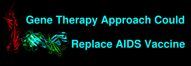
The title figure shows murine 2D5 Fab (heavy chain, green; light chain, cyan) bound to human CD-4 (red) modeled using coordinates from protein protein data bank file accession code 4Q6I from (4).
References
- Gardner, M. R., Kattenhorn, L. M., Kondur, H. R., von Schaewen, M., Dorfman, T., Chiang, J. J., Haworth, K. G., Decker, J. M., Alpert, M. D., Bailey, C. C., Neale, E. S., Jr, Fellinger, C. H., Joshi, V. R., Fuchs, S. P., Martinez-Navio, J. M., Quinlan, B. D., Yao, A. Y., Mouquet, H., Gorman, J., Zhang, B., Poignard, P., Nussenzweig, M. C., Burton, D. R., Kwong, P. D., Piatak, M. Jr, Lifson, J. D., Gao, G., Desrosiers, R. C., Evans, D. T., Hahn, B. H., Ploss, A., Cannon, P. M., Seaman, M. S., Farzan, M. (2015) AAV-expressed eCD4-Ig provides durable protection from multiple SHIV challenges. Nature. 519(7541):87-91. doi: 10.1038/nature14264
- Haigwood, N. L. (2015) HIV: tied down by its own receptor. Nature 519(7541):36-7. doi: 10.1038/nature14205
- Choe, H., Li, W., Wright, P. L., Vasilieva, N., Venturi, M., Huang, C. C., Grundner, C., Dorfman, T., Zwick, M. B., Wang, L., Rosenberg, E. S., Kwong, P. D., Burton, D. R., Robinson, J. E., Sodroski, J. G., and Farzan, M. (2003) Tyrosine sulfation of human antibodies contributes to recognition of the CCR5 binding region of HIV-1 gp120. Cell 114(2):161-70. doi: http://dx.doi.org/10.1016/S0092-8674(03)00508-7
- Pegu, A., Yang, Z. Y, Boyington, J. C., Wu, L., Ko, S. Y., Schmidt, S. D., McKee, K., Kong, W. P., Shi, W., Chen, X., Todd, J. P., Letvin, N. L., Huang, J., Nason, M. C., Hoxie, J. A., Kwong, P. D., Connors, M., Rao, S. S., Mascola, J. R., Nabel, G. J. (2014) Sci. Transl. Med. 6(243):243ra88. doi: 10.1126/scitranslmed.3008992